Wrinkled Retina
 A wrinkle in the retina is a very common problem among older adults. In order to understand what causes this problem and how it can be cured, it is important to learn about the structure of the eye.
The eye is shaped like a small egg and measures about on inch across. The cornea and lens, which lie in the front part of the eye, focus incoming light to form an image on the retina. The retina lines the back wall of the eye and, like the film in a camera, functions to capture and to transmit images to the brain. The central area of the retina where the light is sharply focused is called the macula. The macula allows 20/20 vision and is responsible for reading and other tasks that require fine discrimination. Between the lens of the eye and the retina is a large round space called the vitreous cavity. This space takes up 80% of the volume of the eye and is filled with a gel called the vitreous humor. This gel is normally transparent and is attached to the inner surface of the retina.
Until a person is about 30 years of age, the vitreous gel has the consistency of raw egg whites. After age 30, the gel undergoes a slow degeneration during which pockets of salt water form inside the gel and it starts to sag. Eventually, so much water forms inside the gel that it cannot support itself anymore and the structure collapses. The collapse of the vitreous gel is called a vitreous detachment. When the vitreous detaches, it can cause, among other things, microscopic damage to the inner surface of the retina, especially in the macula. The retina heals itself by the migration and proliferation of certain cells that normally are found within the retina. In most people, the healing process occurs without any further problems. In some people, the healing process goes “haywire” and results in the formation of scar along the inner surface of the retina.
This scar tissue is called an epiretinal membrane. Once the scar tissue forms, several things can happen. In most people, it grows very slowly and eventually stops. In some patients, the scar tissue continues to grow and begins to pull on the retina. When the scar tissue pulls on the retina, the retina becomes wrinkled. So instead of the retina having a smooth surface like the bottom of a glass bowl, it looks like a rippled potato chip. Epiretinal membranes have several synonyms; cellophane membranes, surface wrinkling retinopathy, macular pucker, or simply, a wrinkle in the retina.
Symptoms of a Wrinkled Retina
 The symptoms that you may experience with a wrinkled retina include blurry vision while reading or driving or distortion of lines that are supposed to be straight. A retina specialist can diagnose a wrinkled retina during a detailed, painless examination of the retina using special lenses and instruments.
In some patients, the doctor will recommend a fluorescein angiogram test to determine whether the scar tissue is distorting the retinal blood vessels so much that fluid leaks from these vessels into the retina. This condition is then called macular edema and poses a more serious threat to vision than if no fluid is present.
What will happen to your vision if you have a wrinkled retina? Fortunately, in most patients, vision is only mildly affected. However, in some patients, the vision can decrease to the level of legal blindness over many months. A wrinkled retina will almost never cause total blindness. Wrinkled retinas can affect both eyes in about 1 out of 10 patients, although one eye is usually worse than the other and both eyes are not usually affected at the same time.
Monitoring your Vision
The best way to monitor your vision is to test your eyesight every week with the Amsler Grid that you are given. The treatment that Dr. Kirsch, Dr. Hairston, and/or Dr. Ho recommend for wrinkled retinas is custom designed for your individual situation. If the vision is still good enough for you to read and drive, and the symptoms of distortion are not bothering you very much, conservative observation with very careful regular examinations every 4 to 6 months is usually recommended. The exception to this rule is if there is a significant amount of fluid in the retina found on examination or with the fluorescein dye test. If the symptoms are difficult for you to tolerate or the vision has significantly decreased, then surgery is usually offered as the treatment of choice.
Surgery for Wrinkled Retina
 Before discussing surgery, it is important to understand that there are no medications, eye drops, vitamins, or laser treatments that can treat a wrinkled retina. The operation for a wrinkled retina is performed in the regular operation room and can be done under local or general anesthesia. You may go home the same day or stay overnight in the hospital.
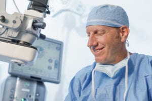
The risks of this operation include retinal detachment (occasionally), infection inside the eye (rarely), and bleeding inside the eye (rarely). These complications are treatable most of the time, although as with any surgery inside the eye, loss of eyesight can rarely result. In some patients (about 1%) the scar tissue grows back. If you already have a cataract, surgery will most likely make it worse. The surgery is called a vitrectomy because the vitreous gel inside the eye is removed through 3 small incisions. Each incision is about 1 millimeter in length and is closed with very fine absorbable sutures. The vitreous gel is replaced with salt water (the vitreous is like your appendix; you have it but you are sometimes better off without it). The edge of the scar tissue is then identified and separated from the surface of the retina. Using special microscopic forceps, the scar tissue is peeled from the surface of the retina like you would peel a label from its backing.
The surgery for wrinkled retinas is successful in the vast majority of patients, although it can take from 6 to 12 months for the vision to improve. The vision and symptoms in eyes with wrinkled retinas can be improved in 80% to 90% of cases. Working with Dr. Kirsch, Dr. Hairston, and/or Dr. Ho, you can decide which course of action is best for your situation to best preserve or restore the precious gift of sight.
Schedule your Retina Evaluation today
Call (727) 581-8706 to schedule your appointment
Meet Your Retina Care Specialists
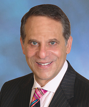
 Leonard S. Kirsch, MD is a fellowship-trained vitreous and retina specialist. He is internationally known for the numerous papers and lectures he has presented here and abroad. Dr. Kirsch is currently an active participant in ongoing research to find new treatments for diseases of the retina. His expertise in the most advanced diagnostic and treatment techniques of all diseases of the retina, macula, and vitreous make Dr. Kirsch one of the elites in his field. In the Tampa Bay area, Dr. Kirsch pioneered the use of Photodynamic Therapy with Visudyne©, and intravitreal Macugen©, Lucentis©, and Avastin© for the treatment of age-related macular degeneration. Dr. Kirsch was also among the first surgeons in Florida to perform 25 gauge “no-stitch” vitrectomy in 2001. He is certified by the American Board of Ophthalmology and is a fellow of the Royal College of Surgeons of Canada.
 Richard J. Hairston, M.D. is a vitreous and retina specialist. He joined The Eye Institute in June 2001 coming to us from a busy retina practice in Washington, DC. Dr. Hairston graduated from the Johns Hopkins University School of Medicine and did his residency at the Wilmer Ophthalmological Institute at Johns Hopkins University. He completed a fellowship in diseases and surgery of the retina and vitreous at The Center for Retina Vitreous Surgery, Memphis, Tennessee. He then served as Assistant Chief of Service in Ophthalmology and Director of the Ocular Trauma Service at Johns Hopkins Hospital. Most recently he was Assistant Professor of Ophthalmology at Johns Hopkins University. He is certified by the American Board of Ophthalmology. Dr. Hairston enjoys an international reputation as an outstanding retina and vitreous surgeon.
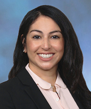
 Janie Ho M.D., is a board-certified ophthalmologist, fellowship-trained in medical and surgical vitreoretinal diseases such as macular degeneration, diabetic retinopathy and retinal tears and detachments. She has been educated at some of America’s finest institutions. She received her Bachelor of Arts from Harvard University and her medical degree from Duke University. She went on to ophthalmology residency at the University of California, San Francisco. Following residency, Dr. Ho continued on to a fellowship in vitreoretinal diseases at the prestigious Massachusetts Eye and Ear Infirmary of Harvard Medical School. Dr. Ho has participated in angiogenesis research, investigating causes and treatment for common retinal disorders.
 Sejal Shah M.D., is a board-certified, fellowship-trained Retina Specialist. Dr. Shah received her medical degree from the University of South Florida. She began her postgraduate training with an internship in medicine at UCLA, followed by ophthalmology training at the University of South Florida. She went on to complete a fellowship at the prestigious Bascom Palmer Eye Institute, specializing in the diagnosis and treatment of retinal disorders. Dr. Shah also earned a B.S. degree with honors in Nutritional Science from the University of Florida. Dr. Shah brings her expertise as a medical retinal specialist with skills in managing and treating vitreoretinal pathology which including age-related macular degeneration and diabetic retinopathy. Her research has appeared in publications such as Ophthalmic Surgery, Lasers, and Imaging Retina; Clinical Ocular Oncology; Survey of Ophthalmology; International Ophthalmology Clinics; and Survey of Ophthalmology. In her spare time, she enjoys spending quality time with her family and friends, traveling, and trying to keep up with her twin boys.

